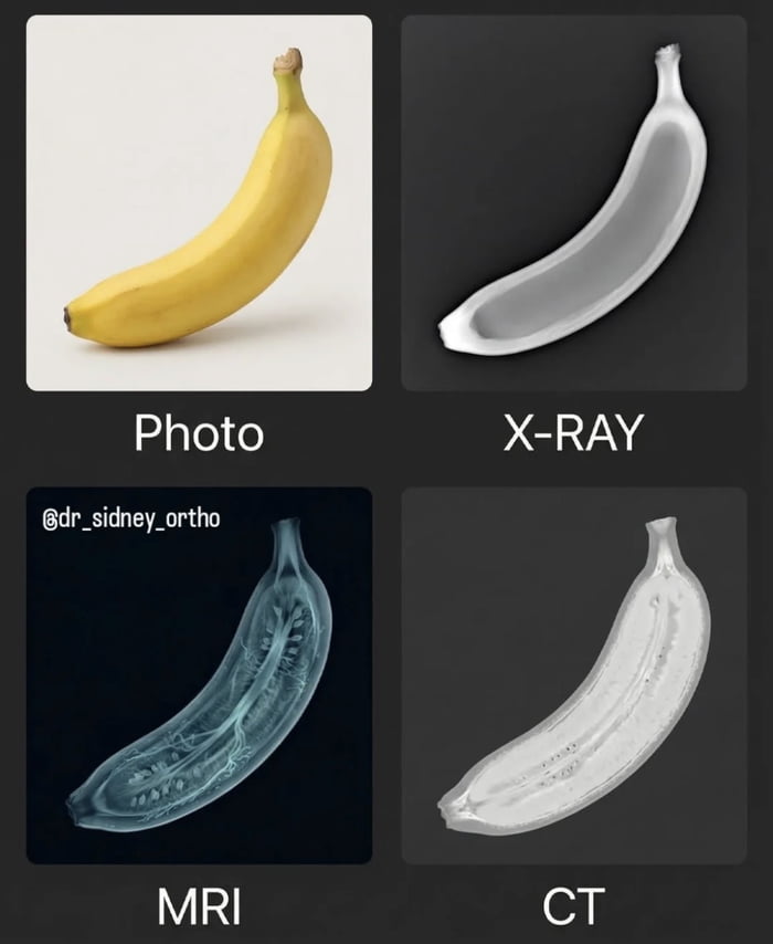
Banana Scanned: Photo vs X-RAY vs MRI vs CT

Text content
Labels: 'Photo', 'X-RAY', 'MRI', 'CT'; Watermark: '@dr_sidney_ortho'
Overview
A 2x2 grid image comparing a banana under different medical imaging techniques. Top-left: Color photo of a ripe yellow banana with a curved shape and greenish stem. Top-right: Labeled 'X-RAY', showing a grayscale outline of the banana's external structure. Bottom-left: Labeled 'MRI', featuring a blue-tinted scan revealing internal details like seeds and fibrous structures. Bottom-right: Labeled 'CT', displaying a high-contrast grayscale cross-section of the banana's interior. A watermark '@dr_sidney_ortho' is positioned in the top-left corner of the MRI quadrant. The image humorously applies professional medical imaging modalities to a common fruit, blending education with lightheartedness.
Origin notes
Originates from the platform 9Gag, as indicated by the provided source information. The image appears to be a user-created collage combining four distinct scans of a banana. The watermark '@dr_sidney_ortho' suggests the medical scan images may have been sourced or created by a user with that handle, possibly in a medical or imaging-related field. The collage method involves arranging four separate images into a grid with descriptive labels, likely intended for educational or humorous sharing on social media.


