This Looks Painful: Spiky Bone Tumor X-Ray vs Actual Specimen
Overview
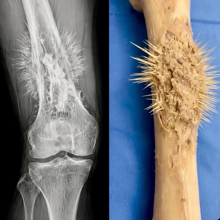
The image presents a side-by-side comparison of two views of an osteosarcoma (bone cancer) lesion on a human femur (thigh bone). The left panel is a medical X-ray showing the femur near the knee joint, with a characteristic 'sunburst' pattern of sharp, spiky abnormal bone growth extending outward from the main bone surface. The right panel is a photograph of an actual excised bone specimen, showing the same type of sharp, spiky, porous abnormal growth protruding from the bone, matching the appearance seen in the X-ray. The spiky growth would severely irritate surrounding soft tissue, leading to extreme pain, as referenced in the original post title.
Origin notes
This post was originally shared on the social media platform 9Gag with the title 'This looks painful'. It combines two public domain medical images: an X-ray of an osteosarcoma lesion and a corresponding surgical specimen photo, assembled into a side-by-side comparison to show the real-world appearance of the abnormal growth seen in the X-ray. It circulated as a shocking, cringeworthy medical content post due to its visceral, painful-looking visual.


